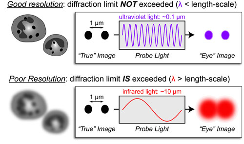
Basic light microscopy can only resolve objects that are larger than 100 nm which means that while it can visualize animal cells (~10,000nm), organelles and bacteria (~1,000nm), it cannot visualize viruses (<100nm), proteins (<10nm) or small molecules(~1nm) (see post summarizing Biological Scales). This limitation is known as the “diffraction limit” and is caused by the fact light only interacts differently with objects separated by more than one wavelength (λ). Intuitively, its helpful to think of each of these wavelengths as “a minimum pixel size” for a computer image where: infrared light (λ ~ 10.0μm) has pixels 100 times larger than ultraviolet light (λ ~ 0.1μm).
For this reason, radar uses microwaves ( λ ~ 1m) to visualize planes and crystallography uses x-rays (λ ~ 1 nm) to visualize small molecules. You can find more examples of the range of microscopic techniques in our post on the Spectrum of Microscopy and Spectroscopy.
REFERENCES:
- Abbe, E. Beitrage zur Theorie des Mikroskops und der mikroskopischen Wahrmehmung. Archiv für Mikroskopische Anatomie 1873, 9, 413–420.
- Lakowicz, J. R. in Principles of Fluorescence Spectroscopy, Springer, 2006
- McQuarrie, D. A.; Simon, J. D. Physical Chemistry: A Molecular Approach., University Science Books, 1999
- Skoog, D. A., Holler, F. J. & Nieman, T. A. Principles of Instrumental Analysis (5th ed), Saunders College Publishing, 1998

This work by Eugene Douglass and Chad Miller is licensed under a Creative Commons Attribution-NonCommercial-ShareAlike 3.0 Unported License.