Category Archives: Cellular Biology

How do you “Fractionate” a Cell?

cell-fractionation-v19

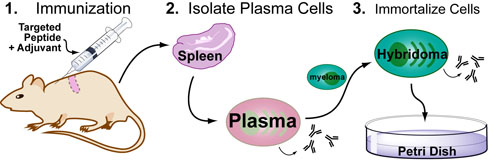
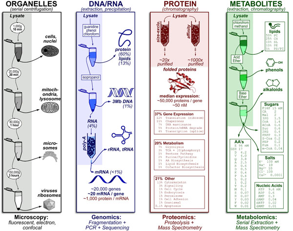
Different scientific questions focus on different parts of the cell and it is often necessary to break a cell up into those different pieces (figure above). While various “-omic” methods are well suited to answering global/systems-level questions for the four catagories listed above (e.g. microscopy, genomics, proteomics, metabolomics) they often lack the resolution of fractionation-methods to answer molecular level questions.

Continue reading

Introduction to Super-resolution Microscopy

microscopy-defraction-limit-v4

While microscopic methods with protein and atom scale resolution – that don’t break the diffraction barrier! – exist (e.g. electron microscopy, atomic force microscopy, x-ray crystallography, etc.), they tend to be more practically difficult than fluorescence-based microscopy (see posts on Fluorescent Probes and Fluorescent Antibodies). Therefore, to give fluorescence microscopy nanometer resolution, several super resolution techniques have been developed that combine clever (1) optical/photophysical and (2) computational-processing tricks to “clean up” the “blurred data.”

Continue reading

Understanding the Diffraction Limit in Microscopy

microscopy-defraction-limit-v4

Basic light microscopy can only resolve objects that are larger than 100 nm which means that while it can visualize animal cells (~10,000nm), organelles and bacteria (~1,000nm), it cannot visualize viruses (<100nm), proteins (<10nm) or small molecules(~1nm) (see post summarizing Biological Scales). This limitation is known as the “diffraction limit” and is caused by the fact light only interacts differently with objects separated by more than one wavelength (λ). Intuitively, its helpful to think of each of these wavelengths as “a minimum pixel size” for a computer image where: infrared light (λ ~ 10.0μm) has pixels 100 times larger than ultraviolet light (λ ~ 0.1μm).

Continue reading

Introduction to ELISA

sandwich-elisa-intro-v6

ELISA or Enzyme-Linked Immunosorbent assay is the most commonly used method for measuring proteins concentrations in solution. It is extensively used both in the laboratory (e.g. culture supernatant) and the clinic (e.g. blood tests) due to its simplicity and adaptability to other protein-based assays such as high throughput screening. The way it works is outlined in the figure above and described in detail below:

Continue reading

Introduction to Flow Cytometry

Web

Flow cytometry is a technique that can quantitatively measure (1) protein expression levels per single cell and (2) amounts of different cell-types (based on a protein-maker) for thousands of cells in minutes! Flow cytometers use hydrodynamic focusing to force a mixture of stained cells into a single-file line through a flow cell. Then, each cell, sequentially, passes through a laser beam which excites the fluorophore allowing quantification of protein expression for all proteins that are stained!

Continue reading

Hybridomas for Large-scale Antibody Production

antibody-production-v3

Following up on our post introducing antibody-based experiments, we wanted to describe how antibodies are made in large volume: First, an animal is immunized with the protein (or peptide) of interest; Second, the spleen is dissected and the plasma cells producing the antibodies of interested are isolated; Finally, these plasma cells are fused with myeloma cells to “immortalize” them into a hybridoma (an antibody-producing cell-line that can be cultured indefinitely).

Continue reading

Introduction to Antibody Based Experiments

Web

Antibodies are immune proteins that can be easily engineered to bind (and “detect”) any given protein in via a simple vaccination method. To “signal” the presence of detected proteins, fluorophores are attached to “stain” the protein (and cell or tissue expressing it) a particular color. These fluorescent probes, have been utilized to identify the location of specific proteins in tissue sections (histology), single cells (microscopy) and quantify the amount of protein in cells (flow cytometry) and in complex mixtures (western blotting).

Continue reading